Intra-operative Brain Deformation
|
|
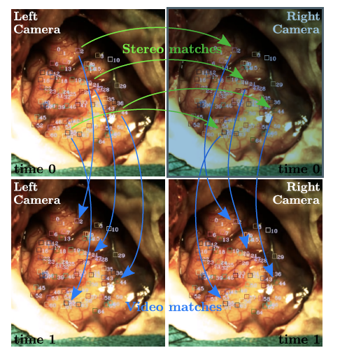
| Real-time brain deformations estimation: Stereo and video feature matching. Each row shows a video frame, and columns stand for the left and right cameras. Stereo matches are depicted by green arrows from the left to the right camera at time 0, and blue arrows show video matches for both cameras from time 0 to time 1. Each feature neighbourhood is depicted as a small numbered square. |
|
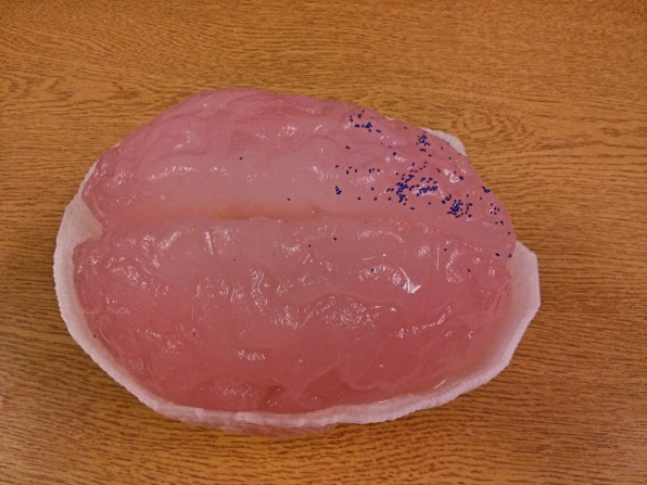
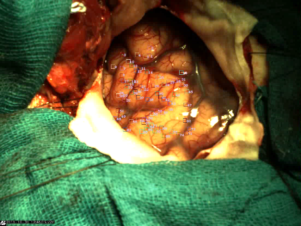
(a) |
(b) |
(c) |
| Experimental setup of a deformable brain phantom for real-time modeling of intra-operative brain shift based on video tracking: (a) The whole phantom; (b) view through a mock craniotomy; (c) tracked features on a live brain. | ||

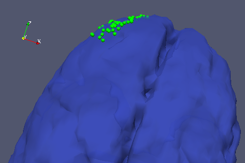
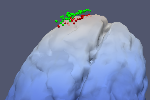
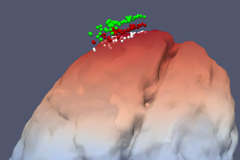
|
(a) |
(b) |
(c) |
|
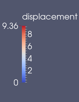
| Results of the brain shift simulation (using the open-source ASL) based on tracked feature points and the corresponding deformation of the brain model, using a 4×4×4 mm3 cell-grid: (a) green points: initially registered to the surface of a brain phantom; (b) Top right, red points: after 500 seconds of deformation tracking; (c) white points: after 1000 seconds of deformation tracking. | |||